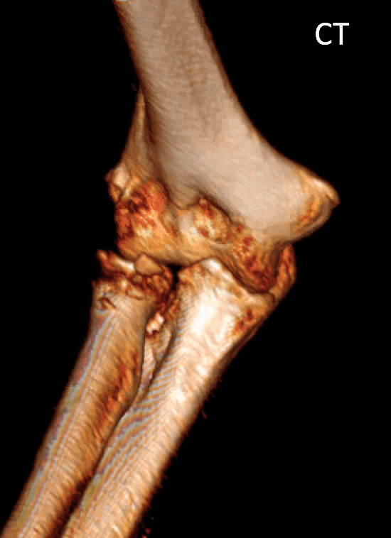
This 59 you female fell sustaining a highly comminuted radial head fracture and non-displaced coronoid fracture. This CT reconstruction reveals the extent of the injury to the radial head. Based on these images, as well as the appearance of the fracture after surgical exposure, the decision was made to replace the bone rather than reconstruct it with plates and screws.

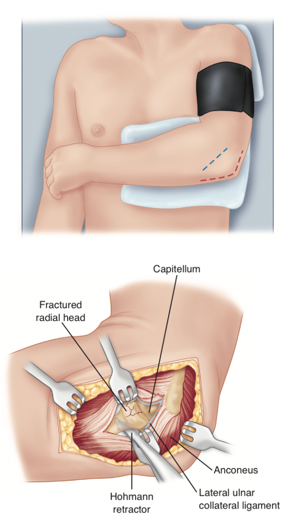
A Lateral incision was made (blue line) and the joint and fracture precisely exposed.


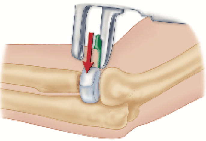
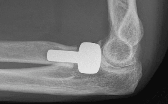
A small saw was used to remove the fractured radial head and neck. After the neck was prepared, trials were used to determine the correct implant size. Next, the final implant is place. The patient began motion exercises just a few days after surgery and recovered with full, painless motion.