This 16-year-old male sustained a fracture at the base of his thumb metacarpal that was treated by another orthopedic physician. The fracture healed in a malunited position that resulted in joint irregularity and painful instability with any thumb use.
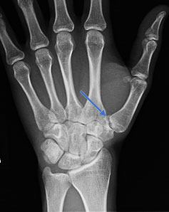
This fracture at the base of the thumb metacarpal bone (Bennett’s Fracture) healed in a poor position as seen on this pre-operative X-ray.
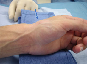
At rest, the thumb metacarpal bone is partially dislocated at the carpometacarpal joint. The arrow highlights the prominence of the dislocated metacarpal base.
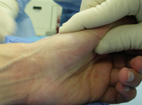
The dislocation is manually reduced with traction and gentle pressure over the boney prominence. When the pressure is released the bone immediately dislocates revealing the instability.
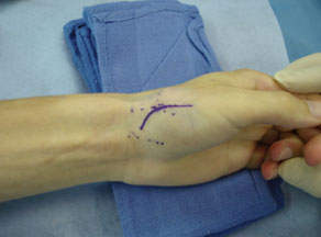
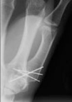
The thumb carpometacarpal joint was exposed and the malunited fracture identified. An osteotome is utilized to mobilize the fracture fragment. The fragment is then anatomically re-positioned to restore the joint congruity and stability. The fracture is stabilized with metal pins (K-wires) which are removed several weeks later after healing has taken place. This high school student returned to all sports/leisure activities without pain or restriction.

High impact professional athlete with wrist pain affecting function and performance. His subtle MRI findings…
Professional golfer with persistent pain over the small finger side (ulnar-sided) of his wrist preventing…

This is a professional basketball player who fell on the court and had immediate wrist…