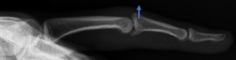
37-year-old businessman who sustained a fracture dislocation of this long finger proximal interphalangeal joint 2.5 months earlier while playing catch with his son. The fracture dislocation was not recognized and healed in a malunited position which then allowed the joint to dislocate dorsally (away from the palm). The patient complained of severe stiffness, swelling and pain with attempts at motion.
This lateral X-ray reveals the multi-fragmented nature of the fracture at the palmar base of the middle phalanx with the resulting dorsal dislocation (arrow) of that bone at the proximal interphalangeal joint (PIP joint) (wrist to the left and fingertip to the right)
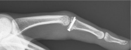
The osteocartilagenous (bone along with a cartilage joint surface) is harvested from a donor site in the wrist, contoured to fit the middle phalanx defect exactly, then secured with K-wires which are exchanged for two screws. The lateral X-ray reveals reduction of the joint and a reconstituted joint surface.
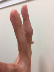
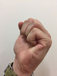
Three months after surgery virtually full motion is restored, the joint is stable and the patient is pain free.

High impact professional athlete with wrist pain affecting function and performance. His subtle MRI findings…
Professional golfer with persistent pain over the small finger side (ulnar-sided) of his wrist preventing…

This is a professional basketball player who fell on the court and had immediate wrist…